
Our research focuses on understanding the cellular and molecular processes that underlie skeletal development and repair in mammals.
Skeletal development
 Our laboratory studies how the skeleton develops in the mammalian embryo, with a particular focus on the formation of the limbs and axial skeleton. To advance our understanding of these processes, we use conditional knock-out techniques.
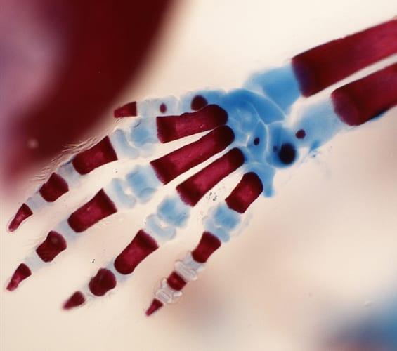
Handplate development
Currently, we are studying a specific mutation that leads to split hand/foot malformation, a condition in which the central digits of the hand or foot are absent. From our studies, we are able to construct a gene regulator network that patterns the digits.
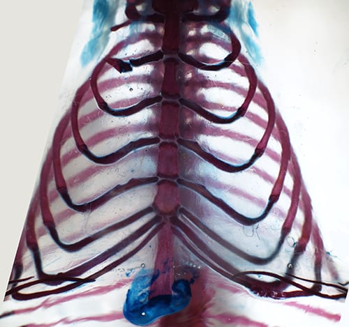
Rib development
Aside from the limbs, another feature of land animals is an enclosed rib cage. Our lab seeks to identify the genes that control rib development and patterning.
We are also exploring the similarities between these underlying mechanisms of development and the underlying mechanisms of repair. By identifying and understanding the connections between these fundamental processes, we hope to inform future cell-based therapies for patients with a wide variety of skeletal injuries and diseases.
Skeletal repair
 Our lab uses conditional knock-out techniques in mouse models and mouse embryonic stem (ES) cells to study bone and cartilage regeneration, focusing on the source of stem cells, symmetric versus asymmetric cell division and cell-cell communication between osteoblasts and osteoclasts. We study situations such as fracture repair where bone heals well, as well as critical-sized injuries where the skeleton or cranium fails to repair. In addition, we have developed a new model for large-scale repair in mammals. It turns out that both the cartilage and the bone portion of the ribs can readily repair in mice and in humans, and we are using this model to understand the cellular and molecular basis of repair. Our studies have implications for ameliorating skeletal injury, chronic osteoarthritis and the severe problems associated with reconstructive surgery.
Source of stem cells
We study the important role of the surrounding connective tissues (periosteum and perichondrium) in housing the cells needed for healing cartilage and bone in mouse ribs. For example, when we remove both a section of rib cartilage and its surrounding sheath of perichondrium, the missing sections fail to repair. However, when we remove rib cartilage but leave its perichondrium, the missing sections entirely repair within one to two months. The perichondrium retains the ability to produce cartilage even when disconnected from the rib and displaced into nearby muscle tissue — further suggesting that the perichondrium contains progenitor or stem cells.
Symmetric versus asymmetric cell division
We use embryonic stem (ES) cells to explore the network of genes, including atypical protein kinase C iota (PRKCI), that control symmetric versus asymmetric cell division. By knocking out the PRCKI gene, we hope to encourage stem cells to favor symmetric cell division, thereby amplifying that population. This could eventually lead to the development of drugs that inhibit the Prcki protein, boosting stem cell production and aiding in skeletal repair.
Cell-cell communication between osteoblasts and osteoclasts
We have identified a previously unknown intracellular characteristic of pluripotent cells, including osteoclasts and osteoblasts: unusually large lysosomes. Under specific conditions, these lysosomes perform exocytosis, secreting molecules that could be important for cell-cell communication. We are studying these lysosomes in the hopes of better understanding how osteoblasts and osteoclasts communicate with each other and maintain the delicate balance between bone formation and resorption.